Leaf hairs and molecules: Microscopy winners of the 2017 NC State Research Image Contest

First place for microscopy among faculty and staff goes to Rich Spontak, Distinguished Professor of Chemical and Biomolecular Engineering, for an image he titled “The Fingerprint of Molecules” (pictured above).
“This unedited polarized light microscope image shows the crystal formation of a special type of polyhedral oligomeric silsesquioxane (POSS) solvent-casted with chloroform,” Spontak says. “We observe plaque-like formations surrounding a fingerprint-like domain, which we found was characteristic only with this molecule and this particular solvent.
“By using computer simulations, we also understood the underlying reasons behind these formations,” Spontak says. “There is a unique molecular and geometric interaction between these molecules. They lock each other just like LEGO pieces. This way, POSS molecules can form a continuous surface that can be used as a protective coating on polymers against X-rays and UV light. We observe here the fingerprint of that perfect protection.”
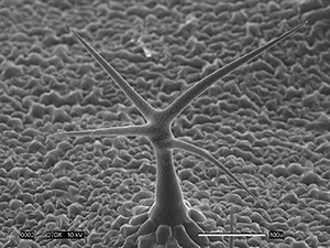
The first place winner for graduate students and postdocs is Eric Land, a Ph.D. student in the Department of Plant & Microbial Biology, for his image of a leaf structure in Arabidopsis thaliana, a model organism widely used in plant biology research.
“This scanning electron micrograph captures the impressive three dimensional structure of an Arabidopsis trichome, or ‘leaf hair,’” Land says. “Appearing like a tree growing among a meadow, these trichomes are actually single cell appendages growing out of the leaf surface. In the background, surface cells, and pore-like stomata can also be seen.
“Trichomes are unique structures, often capable of secreting beneficial compounds onto a plant’s leaf surface,” Land explains. “Investigating how these compounds are coated onto the leaf surface, and what their natural roles are, may provide significant insight into crop resistance to insects, fungi and drought.”
Second place in the grad student category was awarded to Gabrielle Corradino, a Ph.D. student in the Department of Marine, Earth and Atmospheric Sciences, for the image she titled “Nano-Hitchhikers.”
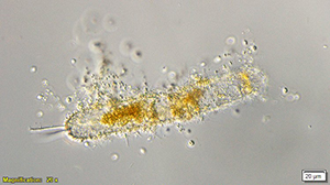
“Nanoflagellates are single-celled planktonic eukaryotes ranging in size from two to 20 microns and can have various nutrient strategies,” Corradino says. “These organisms (not sharks), may be considered the mightiest predators in the ocean. In this image we see over 30 nanoflagellates ‘hitching’ a ride from a larger phytoplankton cell. In some cases, nanoflagellates can infect plankton in marine waters, which plays a significant role in energy transfer and nutrient cycling through marine food-webs.
“Nanoflagellates remain one of the least-characterized members of the marine microbial food-web,” Corradino says. “Understanding their species-specific physiology and trophic interactions is necessary as we aim to forecast energy flow and potential plankton regime shifts in response to anthropogenic stressors in our oceans.”
There was no second-place winner for faculty and staff in the microscopy category.
Note: You can find the work from winners in all of the research image contest categories here.
This post was originally published in NC State News.
- Categories:
